4.1.3. Diospyros abyssinica
The bark of D. abyssinica is black spotted white in
colour. The sapwood is slightly different from heartwood in colour.

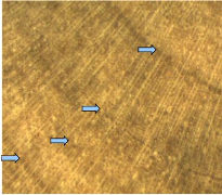
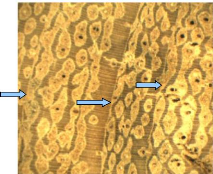
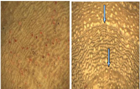
Figure 7: Wood anatomy of D. abyssinica
(Ebenaceae)
The wood colour is usually between white an d yellow and the
heartwood presents black streaks (Figure 7). The identification of tree-rings
is not always easy macroscopically. They are narrow and are presented like
single concentric line. In the growth zones, no vessels were observed. The
boundary zones are characterized by patterns of alternating parenchyma and
fibre bands.
4.1.4. Isoberlinia doka
I. doka tree has moderately furrowed bark whose
colour varies from white to brown. In the studied samples, there were no
significant colour differences between heartwood and sapwood. The wood is
usually brown (Figure 8).

Figure 8: Wood anatomy of I. doka
(Caesalpinaceae)
Tree-rings are darker and wide. They appear like concentric
bands. Thus, macroscopically the boundary zones are distinct and are formed by
tangential lines within a tree ring. In wood cross section, vessels of fairly
uniform are distributed throug hout a growth ring (diffuse-porous). Therefore,
Tree-rings are characterized by marginal parenchyma bands. All vessels are
housed in storage cells (radial parenchyma) that are lighter and show a
parallel disposition to the boundary zone. In each radial par enchyma, we
identified one to three vessels. The radial parenchyma was mostly seen in
sapwood. The Figure 8 shows visible annual ring in I. doka wood stem
disc. In the central core of this stem we noticed a small hole that
demonstrates the unlignified cell walls of pith.
4.1.5. Pterocarpus erinaceus
P. erinaceus bark is deeply furrowed with dark mahogany
in colour. The wood is yellowish and no difference was noticed between sapwood
and heartwood (Figure 9).

Figure 9: Wood anatomy of P. erinaceus
(Fabaceae).
The observed structure through high accuracy showed a
decreasing of vessels size towards the tree-ring. Then, just after one ring,
the vessel is wide and the size decrease s gradually until the next boundary
growth, thus a clear distinction of the rings. The boundary growth has circular
slightly undulating form. The presence of alternating fibres and parenchyma
tissues is remarkable.
4.1.6. Conclusion on wood anatomy
For each investigated species, the wood anatomical structure
showed a variati on from one to another. We also identified three different
tree-ring structures:
> border of rings presenting variation in vessels distr
ibution that was the case of A. leiocarpa (Combretaceae) and P.
erinaceus (Fabaceae) species;
> border of rings delimited by marginal parenchyma bands which
was represented by D. microcarpum (Caesalpiniaceae) and I. doka
(Caesalpiniaceae) species;
> growth ring boundary like alternating bands of fibre and
parenchyma cells illustrated by D. abyssinica (Ebenaceae).
The visual analysis demonstrated that there are differences in
tree-ring structures among the species. Thus, genetic impact is
questionable.
On the other hand, throughout the wood colour, we categorized the
targeted species in three major groups:
> white to yellow with black streaks wood illustrated by
D. abyssinica;
> yellowish wood that regroups A. leiocarpa and
P. erinaceus;
> light brown to dark brown wood that are the case of D.
microcarpum and I. doka. Both species are in the same family of
Caesalpiniaceae .
Finally, about the distinctiveness of ring borders, we
conclude that I. doka, D. microcarpum, and P. erinaceus
have the best distinct rings. As far as the D. abyssinica is
concerned, it showed annual tree-rings but the use of high accuracy method is
necessary.
| 


